Mortons neuroma MRI – Clinical vs. Radiological Diagnosis

What is the best way to diagnose Morton’s neuroma? Is Morton’s neuroma a clinical diagnosis? do I need an MRI for Morton’s neuroma?

Morton neuroma, a fairly common musculoskeletal condition is usually diagnosed clinically by an experienced clinician after a full clinical assessment which involves a history and competent clinical examination. However, even in the hands of an experienced clinician, Morton’s neuroma can be a difficult diagnosis to make.

What should you know about Morton’s Neuroma diagnosis?

The clinical symptoms are usually suggestive of Morton neuroma are(1):

  • Sensation of walking on a pebble/ stone
  • Limitation of motion due to pain and discomfort (especially after activity)
  • Improvement in symptomatology with rest and relaxation

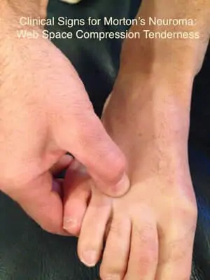
Clinical signs that points to Morton neuroma are:

  • Altered/ decreased sensation in the region of affected nerve
  • Squeezing or pressurizing the forefoot (especially dorsal aspect) may reproduce pain symptoms. Investigators believe that almost all the patients (i.e.100%) experience pain on deep palpation.
foot neuroma
  • On deep palpation, your doctor may feel the enlargement of nerve (or lump); however, this is only observed in cases of significant nerve inflammation/ enlargement
  • Usually no active signs of physical injury or inflammation (such as redness etc.) are seen.
  • Aggravation of pain symptoms is also experienced by patients on weight bearing. According to a new study, this sign is positive in more than 91% patients(1).

Clinical tests with high sensitivity and specificity include:

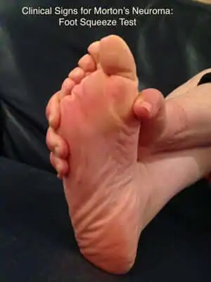
Mulder’s Sign: This test is positive in 98% patients and is usually performed with patient in the lying position. The examiner squeezes the forefoot with one hand while press the ball of the foot with other to produce characteristic pain and often a click (also known as Mulder’s click).  See this Video.

Tinel’s Sign: If the involved space is pressed or squeezed, patient experiences tingling and numbness; which is a sign of nerve inflammation or involvement.

When is radiological diagnosis necessary?

A proper history and thorough clinical examination can help in the identification of most cases of Morton neuroma; but in some cases, it is important to opt for radiological tests to help. These selective situations are:

The diagnosis can remain ambiguous after decent history-taking and examination. This could be due to a number of reasons:

  • Unusual Anatomy. Certain unusual anatomical and physiological characteristics of neuroma (or the foot) may make it challenging for healthcare professionals to precisely identify Morton neuroma.
  • Similar clinical picture. There are many conditions that can mimic Morton’s neuroma.
  • Post surgery or post procedures. Some patients have had numerous invasive procedures done sometimes by relatively inexperienced clinicians and it is difficult to ascertain whether their pain is due to Morton’s neuroma or as a result of the various treatments and procedures that hey have had.
  • Individual foot biomechanics: Morton neuroma is often a result of poor lifestyle and foot-wear choices. Therefore, poor foot dynamics may lead to other foot conditions as well; such as bursitis, ganglion cysts, hammertoe, tarsal tunnel syndrome, arthritis of metatarsal heads or other related injuries.
  • Involvement of more than one web-space: If more than one web-space (or inter-digital space) is involved, it is usually a good idea to opt for radiological investigations; such as ultrasound.

The gold standard for diagnosing Morton’s neuroma is a diagnostic local injection.

Most importantly, Morton’s neuroma is a clinical diagnosis made by a clinical who is experienced in seeing and treating Morton’s neuroma patients. The diagnosis is made a after a careful assessment of the history and physical examination. In some cases a diagnostic injection may be required. It is not a diagnosis made by any radiological test whether that be an MRI or ultrasound.

References:

  1. Pastides, P., El-Sallakh, S., & Charalambides, C. (2012). Morton’s neuroma: a clinical versus radiological diagnosis. Foot and Ankle Surgery, 18(1), 22-24.
  2. Lee, K. S. (2009). Musculoskeletal ultrasound: how to evaluate for Morton’s neuroma. American Journal of Roentgenology, 193(3), W172-W172.
  3. Zanetti, M., Strehle, J. K., Kundert, H. P., Zollinger, H., & Hodler, J. (1999). Morton Neuroma: Effect of MR Imaging Findings on Diagnostic Thinking and Therapeutic Decisions 1. Radiology, 213(2), 583-588.
  4. Resch, S., Stenstrom, A., Jonsson, A., & Jonsson, K. (1994). The diagnostic efficacy of magnetic resonance imaging and ultrasonography in Morton’s neuroma: a radiological-surgical correlation. Foot & Ankle International, 15(2), 88-92.

Get Your Free Morton’s Neuroma Guide:

The top 10 signs you may have Morton’s neuroma and
The top 10 things to do if you think you have Morton’s neuroma

By providing us with your information you are consenting to the collection and use of your information in accordance with our Terms of Service and Privacy Policy.