Here are a few helpful symptoms that may suggest Morton’s neuroma.
It is highly recommended to see your provider for a complete evaluation and diagnosis of Morton’s neuroma. This is mainly because, other musculoskeletal conditions (such as arthritis, nerve damage, traumatic injury etc.) presents with similar signs and symptoms.
It should be noted that a negative Tinel’s sign or Mulder’s sign does not rule out a Morton’s neuroma.
Radiological investigation may be used to confirm the exact site, location and size of the neuroma. Most frequently employed tests are:
X-ray: X-rays can rule out any fracture (especially stress fractures that are fairly common in young girls or athletes). Additionally, X-rays also helps in excluding other injuries or lesion of metatarsals.
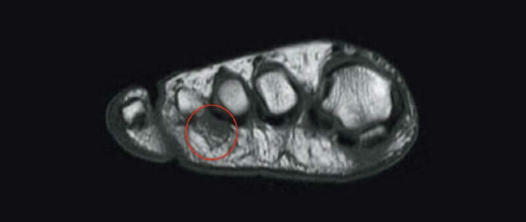
MRI: MRI (magnetic resonance imaging) can be performed to diagnose a Morton’s neuroma. In addition, MRI also helps in excluding other potential causes of pain and discomfort; such as tumor growth or mass, present between the third and fourth toe. However a negative MRI does not exclude the presence of a Morton’s neuroma.
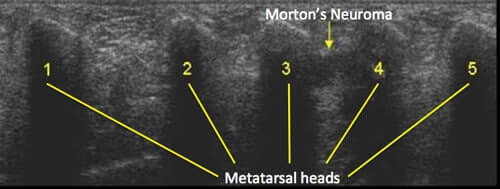
Ultrasound: Ultrasound is a cost-effective radiological modality that is used for identification of exact site and size of Morton’s neuroma. Many practitioners use ultrasound to guide their procedures such as diagnostic injections, cryosurgery, radio frequency ablation and neurolytic injections. According to a study reported in the peer reviewed journal Clinical Radiology(1), the sensitivity of Ultrasound in the diagnosis of Morton’s Neuroma is 90% which is about the same as MRI yet investigators believe that ultrasound is the preferred option over MRI if neuroma is under 5mm in dimensions(2). A negative Ultrasound does not exclude the presence of a Morton’s neuroma.
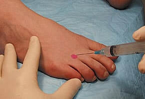
Diagnostic injection is a tool which is frequently used for accurate diagnosis of painful Morton’s neuromas. As part of the procedure, a local anesthetic agent such as lidocaine is injected into the suspected neuroma, preferably under ultrasound guidance. If the pain significantly decreases after introducing the anesthetic agent in the web-space, a diagnosis of Morton’s neuroma is generally made. A diagnostic injection is often considered the gold standard for diagnosis.
Morton’s neuroma is above all a clinical diagnosis. A positive MRI or Ultrasound does not necessarily confirm the diagnosis of Morton’s neuroma. You can have a clear Morton’s neuroma on MRI or ultrasound but the cause of your pain may not be that Morton’s neuroma. It is estimated that up to 60% of the population have Morton’s neuromas on MRI or ultrasound but have no symptoms.
Similarly, a negative MRI or ultrasound does not exclude the diagnosis of Morton’s neuroma. Some Morton’s neuromas are very small and very hard, even impossible to detect with MRI or Ultrasound but may be extremely painful.
It is critical that you see a practitioner well experienced with Morton’s neuroma. For more information on the diagnosis of Morton’s neuroma, see here.

By providing us with your information you are consenting to the collection and use of your information in accordance with our Terms of Service and Privacy Policy.